Lincoln Ave Cat & Dog Hospital
MASS & TUMOR SURGERY
Lump evaluation, biopsy, and surgical removal for dogs, cats, and exotic pets in Bergen County. Diagnosis first, so you know what you're deciding about.
Finding Out What It Is
Most lumps are not cancer. The difference is not something anyone can tell by looking or feeling, which is why the first step is almost always a needle sample rather than a scalpel.
Knowing what a mass is before surgery changes how much tissue needs to come out, whether the chest should be X-rayed first, and sometimes whether surgery is the right choice at all.
35 years of surgical experience
In-house cytology and biopsy
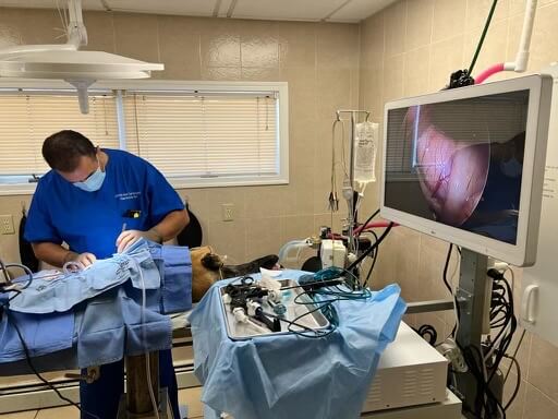
In-house diagnostics and surgery
Dogs, cats and exotic pets
No referral needed
Independent, doctor-owned hospital
Available treatment varies according to the species and condition.
★★★★★ 4.6 · 425+ Google Reviews
Should This Lump Be Checked?
A needle sample takes minutes and usually doesn't require sedation. It's the cheapest, fastest way to stop guessing.
Any new lump you haven't had checked.
A lump that is growing, changing shape or texture.
A lump that has become red, warm, ulcerated or is bleeding.
A lump your pet licks, scratches or reacts to when touched.
You need for a surgical opinion.
In-House Diagnostics and Surgery
Start with an examination and understand the diagnosis before making a treatment decision.
Diagnosis & Surgery in One Hospital
Sampling, imaging and surgery happen in the same building, so a lump found on a Tuesday doesn't require three appointments at three facilities.
Fine needle aspirates and cytology
Surgical and incisional biopsy
Digital X-rays
Ultrasound
Anesthesia monitoring
Bloodwork
Some cytology results may be available the same day. Tissue sent for full pathology takes longer.
What We Diagnose & Remove
Not every mass needs surgery, and not every mass should be removed the same way.
What it is determines what the operation looks like.
Lump Evaluation & Diagnosis
A fine needle aspirate draws a small sample of cells through a needle. Quick, usually no sedation, and often read the same day.
It tells us whether we're looking at a fatty lump, an infection, a cyst or something that needs to come out with margins.
When cytology can't give a clear answer, a biopsy takes a small piece of tissue for full pathology instead.
Fine needle aspirate and cytology
Incisional or punch biopsy when needed
Usually performed in a standard appointment
Determines the surgical plan before surgery is scheduled
Skin & Subcutaneous Masses
Lumps under the skin are the most common thing we're asked about. Lipomas (soft, benign fatty masses) are frequent in older dogs, and many never need removal.
The ones we do remove are those that interfere with movement, sit where a harness or collar rubs, or grow large enough to be uncomfortable.
Cysts, warts, abscesses and skin tags are also removed when they bother the patient or won't resolve on their own.
Lipoma removal when size or location warrants it
Sebaceous cysts and skin tags
Removal of masses that are ulcerated or infected
Tissue submitted for pathology
Mast Cell Tumors & Soft Tissue Sarcomas
These are the reason we aspirate first. A mast cell tumor can look and feel exactly like a harmless fatty lump. It's often called the great pretender, and it's common in Boxers, Bulldogs, Pugs, Boston Terriers, Labradors and Golden Retrievers.
Both mast cell tumors and soft tissue sarcomas extend beyond what you can feel. Removing only the visible lump usually leaves cells behind and the mass regrows. These require a wider excision with a margin of normal tissue around and beneath them.
Wide-margin surgical excision
Reconstructive closure when a large area is removed
Grade guides what, if anything, comes next
Mammary Tumors
Common, and very different between dogs and cats. In dogs, a substantial share of mammary masses are benign, and removal is often straightforward. In cats, mammary tumors are far more likely to be malignant and behave more aggressively, which usually means removing a wider area of tissue rather than a single lump.
Spaying early meaningfully reduces the risk in both species.
Single mass removal through full chain mastectomy
Approach differs substantially between dogs and cats
Chest imaging before surgery in most cases
Spay discussed where relevant
Abdominal Masses
Masses on the spleen or liver often announce themselves by bleeding. Some are found on a routine ultrasound. Others present as a dog who was fine yesterday and collapsed today.
Splenectomy removes the entire spleen, dogs live normally without one. Liver masses may be removed by taking the affected lobe. Both are major abdominal procedures and both are sometimes performed as emergencies.
Splenectomy, planned or emergency
Liver lobectomy
Kidney and adrenal masses
Staging & What Comes After
Surgery is one part of the plan, not always the whole of it. For masses with a tendency to spread, we look before operating ( chest X-rays, abdominal ultrasound, and sampling of nearby lymph nodes) so the surgical plan is built on the full picture.
Chest and abdominal imaging before surgery
Lymph node sampling where indicated
Full histopathology on everything removed
FINANCING OPTIONS WE WORK WITH



Trusted by Fair Lawn Pet Parents
Read how Dr. El-Akbawy and our dedicated team have transformed the lives of pets across Bergen County.
Proud Members & Accreditations:
AAHA Accredited
AVMA Member
NJVMA
Ready to Schedule Your Pet's Visit?
Dogs, cats, and exotics, cared for on Lincoln Avenue Hospital.
Ask for same-day appointments, no referral needed.
Lincoln Ave Cat & Dog Hospital
Family-owned and independently operated on Lincoln Avenue since 1984.


Hours
Mon, Tue, Thu, Fri: 9 - 5pm
Wed: 12pm - 7pm
Sat: 9 - 2pm
Sun: Closed
© 2026 Lincoln Ave Cat & Dog Hospital. All rights reserved. · Privacy · Terms of Service · Accessibility
Serving Fair Lawn, NJ and Bergen county since 1984.